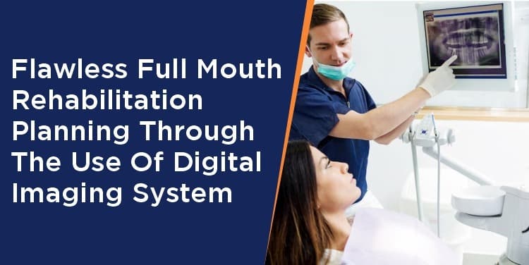
A full mouth reconstruction is one of the most complicated dental procedures to perform, for both the practitioner and the lab technician. The personality and general health of a person can both be significantly improved by a full mouth repair. These kinds of cases are tricky, and success depends on careful planning and assistance.
Digital radiography refers to x-ray imaging. This uses digital x-ray sensors which produce enhanced images of your dental structure on the screen. These radiographs are used to diagnose, discuss and treat any dental condition.

Types of Dental Digital radiography
These digital radiographs can be taken intra-orally or extra-orally. The intra-oral radiographs are mainly used to check for any decay or bone loss concerning teeth whereas extraoral radiography is used to examine teeth, TMJ or any abnormality or fracture if present in the jaw, face or skull.
What are the advantages of Dental digital imaging?

- Digital radiographs reveal a small hidden area of decay with a good resolution to appreciate the caries
- Digital radiographs can be viewed instantly on any computer screen and can be transferred electronically to other dentists or patients for the record.
- Early detection of dental problems can save patients time and money
- Digital radiographs eliminate the chemical processing and disposal of hazardous wastes generated from the traditional ray film used
- Digital sensors require 50-80% less radiation than film
- The contrast, sharpness, and zoom helps in easy appreciation of the defects
After the introduction of the intraoral digital imaging system and the huge success obtained, the extraoral digital imaging system came into existence. This extra oral radiography includes various types like:
- Panoramic x-ray
- Cephalometric projection
- Cone beam computerized tomography
- Multi-slice computer tomography
Panoramic X-ray and Cone beam computed tomography are very frequently used in routine dental practice

Panoramic X-ray: These X-rays help in getting the entire image of the dentition. This is a machine that rotates around the head and shows the entire mouth, including all teeth. These x-rays are beneficial in explaining treatment plans for dental implants, impacted teeth, or any tumour/lesions and bony defects. Also, panoramic x-rays are useful for forensic and legal purposes to identify the unrecognizable foreign object if present.

On the other hand, Cone Beam Computed Tomography (CBCT) shows the body’s interior structure as a 3D image. It is mainly used to identify facial bone problems like tumors or fractures. It can also be used to evaluate the placement of dental implants or in cases of difficult extractions to avoid possible complication
For flawless full mouth rehabilitation or smile design, these two digital imaging are of great help, as an entire picture of teeth, jaws, any defects or foreign body present can be determined. Also, in full-mouth cases where multiple implants are to be placed or during extraction of a third molar checking the proximity of the tooth to the nerve, is of great help.
Genoray OPG & CBCT:

Genoray is one such brand that has both OPG and CBCT machines.
Genoray Papaya 3D Premium Plus is one of the machines used for both OPG and CBCT. It is a combination of, CT, panoramic and cephalometric
There are 3 different dedicated CMOS and amorphous silicon flat panel sensors for different needs. Hence there is long-lasting performance and long life as no overload is there on a single sensor for both 2D and 3D images.
There is no requirement for additional adjustment, thus providing a high-quality scan with the smart position feature which creates a database based on patient-specific exposure and position of the precious image.
There is a smart metal artifact feature called SMARF for more clarity, and scout mode to avoid positioning errors. There is a provision that the image can be captured within 2 sec, thus preventing patient movement and increasing image quality. Also, there is the availability of 1 shot ceph in premium versions. This version of image capability provides the user with accurate information for implant planning, TMJ analysis, airway analysis.
The machine gives accurate results compared to the normal 2D image as it has a variable focal trough with 9 different arch shapes. There is open positioning with 3 scan modes (Normal, Fast, and HD). The machine is self-standing, hence is no requirement for any drill in the wall or floor. It is wheelchair accessible and has a sealed base, so safe from rats and lizards from wire cutting and short circuits.
Conclusion:
Genoray premium plus machine incorporates innovative imaging technology with its unique and more versatile features. This machine is designed to make your work seem easy and accurate for full mouth rehabilitation.